Introduction:
Polyp detection during screening and surveillance colonoscopy has been an important outcome of the study. Despite of thorough detailed examination, smaller polyps can be missed even by a skilled and experienced endoscopist owing to its small size and near similar background and may impact a great cost to the health of patient.
Objective:
To evaluate the effectiveness of recent developments in Endoscopic imaging in terms of AI/CAD ( Artificial Intelligence/Computer Aided Detection) and TXI ( Texture and Color enhancement Imaging) for detection of lesions and increase ADR (Adenoma Detection Rate)
History:
A 65 year old NRI male came for screening colonoscopy with a strong family history of CRC. His routine blood reports were within normal limits. USG whole Abdomen- Normal
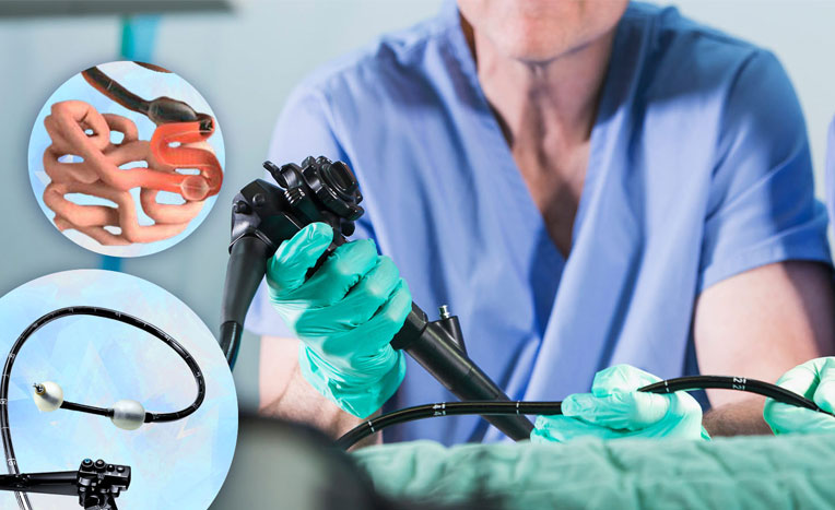
Equipment Used:
EVIS X1 CV-1500, ENDO-AID CADe with CF-EZ 1500 colonoscope on OEV321UH-4K LCD monitor.
Conclusion:
Application of ENDO-AID CADe during examination picked up a very subtle polyp which was unremarkable on conventional white light. After target lesion has been focused further characterization in terms of texture and color enhancement was performed with use of TXI mode I which clearly delineated the lesions. With new generation of scope with higher magnification, features like TXI, AI/CADe, RDI and NBI, it is possible to pick up early malignant/premalignant lesions which are amenable for endoscopic resection.
Images:

Fig. I white light HD

Fig. II ENDO-AID CADe- Lesion detection

Fig. III TXI mode I- Lesion characterization
Dr Chintan Patel
Dept. of Interventional GI Endoscopy,
SIDS Hospital and Research center,
Surat, Gujarat, India.